

Anti-Human Nuclear Antigen Antibody [235-1]
![Immunofluorescence - Anti-Human Nuclear Antigen Antibody [235-1] (A250966) - Antibodies.com](assets/img/products/single-v1/gallery/th01.jpg)
![Flow Cytometry - Anti-Human Nuclear Antigen Antibody [235-1] (A250966) - Antibodies.com](assets/img/products/single-v1/gallery/th02.jpg)
![Immunohistochemistry - Anti-Human Nuclear Antigen Antibody [235-1] (A250966) - Antibodies.com](assets/img/products/single-v1/gallery/th03.jpg)
![SDS-PAGE - Anti-Human Nuclear Antigen Antibody [235-1] (A250966) - Antibodies.com](assets/img/products/single-v1/gallery/th04.jpg)
![Flow Cytometry - Anti-Human Nuclear Antigen Antibody [235-1] (A250966) - Antibodies.com](assets/img/products/single-v1/gallery/th05.jpg)
![Flow Cytometry - Anti-Human Nuclear Antigen Antibody [235-1] (A250966) - Antibodies.com](assets/img/products/single-v1/gallery/th06.jpg)
![Flow Cytometry - Anti-Human Nuclear Antigen Antibody [235-1] (A250966) - Antibodies.com](assets/img/products/single-v1/gallery/th07.jpg)
Anti-Human Nuclear Antigen Antibody [235-1]
Mouse monoclonal [235-1] antibody to Human Nuclear Antigen for Flow Cytometry, IF, IP, ICC and IHC-Fr.
Product size:
Formats and conjugates:
Shipping options:
- Freight / packing charges: £60 to Germany
- Dispatched from: Cambridge (UK)
- Lead time: 5-8 business days
Product details
- Name
- Anti-Human Nuclear Antigen Antibody [235-1]
- Description
- Mouse monoclonal [235-1] antibody to Human Nuclear Antigen.
- Specificity
- This antibody is an excellent marker for human cells in xenographic model research. It reacts specifically with human cells. It is a part of a new panel of reagents, which recognizes subcellular organelles or compartments of human cells. These markers may be useful in identification of these organelles in cells, tissues, and biochemical preparations. MAb 235-1 recognizes an antigen associated with the nuclei in human cells. It can be used to stain the nuclei in cell or tissue preparations and can be used as a nuclear marker in subcellular fractions. It produces a speckled pattern in normal and malignant cells and may be used to stain the nuclei of cells in fixed or frozen tissue sections. It can also be used with paraformaldehyde fixed frozen tissue or cell preparations.
- Applications
- Flow Cytometry, IF, IP, ICC, IHC-Fr
- Dilutions
- Flow Cytometry: 1-2 µg/million cells, IF: 1-2 µg/ml, IP: 1-2 µg / 500 µg protein, ICC: 1-2 µg/ml, IHC-Fr: 1-2 µg/ml
- Reactivity
- Human, Non-Human Primates
- Cross reactivity
- This antibody does not cross react with Mouse, Rat, or Chicken.
- Immunogen
- Nuclei of human myeloid leukemia biopsy cells.
- Host
- Mouse
- Clonality
- Monoclonal
- Clone ID
- 235-1
- Isotype
- IgG1
- Light chains
- kappa
- Conjugate
- Unconjugated
- Purification
- Protein A/G chromatography.
- Concentration
- 200 µg/ml
- Molecular weight
- 70 kDa / 80 kDa
- Product form
- Liquid
- Formulation
- Supplied in 10mM Phosphate Buffered Saline with 0.05% BSA and 0.05% Sodium Azide.
- Storage
- Shipped at 4°C. Upon delivery aliquot and store at -20°C. Avoid freeze / thaw cycles.
Scientific validation data (7)
![Immunofluorescence - Anti-Human Nuclear Antigen Antibody [235-1] (A250966) - Antibodies.com](assets/img/products/single-v1/gallery/01.jpg)
Immunofluorescence - Anti-Human Nuclear Antigen Antibody [235-1] (A250966)
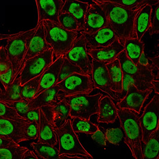
Immunofluorescent analysis of paraformaldehyde fixed HeLa cells stained with Anti-Human Nuclear Antigen Antibody [235-1] followed by Goat Anti-Mouse IgG (CF® 488) (Green). Counterstain is Phalloidin (Red).
![Flow Cytometry - Anti-Human Nuclear Antigen Antibody [235-1] (A250966) - Antibodies.com](assets/img/products/single-v1/gallery/02.jpg)
Flow Cytometry - Anti-Human Nuclear Antigen Antibody [235-1] (A250966)
Flow cytometric analysis of paraformaldehyde fixed HeLa cells using Anti-Human Nuclear Antigen Antibody [235-1] followed by Goat Anti-Mouse IgG (CF® 488) (Blue). Isotype Control (Red).
![Immunohistochemistry - Anti-Human Nuclear Antigen Antibody [235-1] (A250966) - Antibodies.com](assets/img/products/single-v1/gallery/03.jpg)
Immunohistochemistry - Anti-Human Nuclear Antigen Antibody [235-1] (A250966)
Immunohistochemical analysis of acetone-fixed frozen human tonsil using Anti-Human Nuclear Antigen Antibody [235-1].
![SDS-PAGE - Anti-Human Nuclear Antigen Antibody [235-1] (A250966) - Antibodies.com](assets/img/products/single-v1/gallery/04.jpg)
SDS-PAGE - Anti-Human Nuclear Antigen Antibody [235-1] (A250966)
SDS-PAGE analysis of Anti-Human Nuclear Antigen Antibody [235-1] under non-reduced and reduced conditions; showing intact IgG and intact heavy and light chains, respectively. SDS-PAGE analysis confirms the integrity and purity of the antibody.
![Flow Cytometry - Anti-Human Nuclear Antigen Antibody [235-1] (A250966) - Antibodies.com](assets/img/products/single-v1/gallery/05.jpg)
Flow Cytometry - Anti-Human Nuclear Antigen Antibody [235-1] (A250966)
Flow cytometric analysis of PFA fixed Jurkat cells using Anti-Human Nuclear Antigen Antibody [235-1] followed by Goat Anti-Mouse IgG (CF® 488) (Blue). Isotype Control (Red).
![Flow Cytometry - Anti-Human Nuclear Antigen Antibody [235-1] (A250966) - Antibodies.com](assets/img/products/single-v1/gallery/06.jpg)
Flow Cytometry - Anti-Human Nuclear Antigen Antibody [235-1] (A250966)
Flow cytometric analysis of PFA fixed K562 cells using Anti-Human Nuclear Antigen Antibody [235-1] followed by Goat Anti-Mouse IgG (CF® 488) (Blue). Isotype Control (Red).
![Flow Cytometry - Anti-Human Nuclear Antigen Antibody [235-1] (A250966) - Antibodies.com](assets/img/products/single-v1/gallery/07.jpg)
Flow Cytometry - Anti-Human Nuclear Antigen Antibody [235-1] (A250966)
Flow cytometric analysis of PFA fixed MCF-7 cells using Anti-Human Nuclear Antigen Antibody [235-1] followed by Goat Anti-Mouse IgG (CF® 488) (Blue). Isotype Control (Red).
Product citations (4)
Reduced neural progenitor cell count and cortical neurogenesis in guinea pigs congenitally infected with Toxoplasma gondii
- Species: Pig
- Application: Western Blot
- Authors: AR Nanna, AV Kel'in, C Theile, JM Pierson, ZX Voo, A Garg, JK Nair, MA Maier, K Fitzgerald, C Rader.
Reduced neural progenitor cell count and cortical neurogenesis in guinea pigs congenitally infected with Toxoplasma gondii
- Species: Pig
- Application: Western Blot
- Authors: AR Nanna, AV Kel'in, C Theile, JM Pierson, ZX Voo, A Garg, JK Nair, MA Maier, K Fitzgerald, C Rader.
Reduced neural progenitor cell count and cortical neurogenesis in guinea pigs congenitally infected with Toxoplasma gondii
- Species: Pig
- Application: Western Blot
- Authors: AR Nanna, AV Kel'in, C Theile, JM Pierson, ZX Voo, A Garg, JK Nair, MA Maier, K Fitzgerald, C Rader.
Reduced neural progenitor cell count and cortical neurogenesis in guinea pigs congenitally infected with Toxoplasma gondii
- Species: Pig
- Application: Western Blot
- Authors: AR Nanna, AV Kel'in, C Theile, JM Pierson, ZX Voo, A Garg, JK Nair, MA Maier, K Fitzgerald, C Rader.
Reviews (3)
Ryan Murray, University of Leeds
June 25, 2024
- Application: Western Blot, Immunofluorescence
- Species: Human
- Sample: Human aortic endothelial cells (HAECs)
-
Experimental
conditions: WB: 4-20% gel, PVDF membrane, 1:3000 primary. IF: 4% PFA fixation, 0.1% Triton-X permeabilization, primary 1:300, secondary 1:300. - Dilution: WB: 1:3000, IF: 1:300
- Results summary: Observed 95kDa band (CRACR2A-L). High specificity, low background, high sensitivity, reproducible.
- Comments: Minimal background, clear clustering, strong signal.
Ryan Murray, University of Leeds
June 25, 2024
- Application: Western Blot, Immunofluorescence
- Species: Human
- Sample: Human aortic endothelial cells (HAECs)
-
Experimental
conditions: WB: 4-20% gel, PVDF membrane, 1:3000 primary. IF: 4% PFA fixation, 0.1% Triton-X permeabilization, primary 1:300, secondary 1:300. - Dilution: WB: 1:3000, IF: 1:300
- Results summary: Observed 95kDa band (CRACR2A-L). High specificity, low background, high sensitivity, reproducible.
- Comments: Minimal background, clear clustering, strong signal.
Ryan Murray, University of Leeds
June 25, 2024
- Application: Western Blot, Immunofluorescence
- Species: Human
- Sample: Human aortic endothelial cells (HAECs)
-
Experimental
conditions: WB: 4-20% gel, PVDF membrane, 1:3000 primary. IF: 4% PFA fixation, 0.1% Triton-X permeabilization, primary 1:300, secondary 1:300. - Dilution: WB: 1:3000, IF: 1:300
- Results summary: Observed 95kDa band (CRACR2A-L). High specificity, low background, high sensitivity, reproducible.
- Comments: Minimal background, clear clustering, strong signal.
Anti-Human Nuclear Antigen Antibody [235-1] (A250966)
£365
- Product size: 100µg
- Formats & conjugates: FITC
Publishing research using Anti-GFAP Antibody (A85419)?
Let us know, we can list the citation on this page.
Submit
Understanding Secondary Antibodies (Anti-Rabbit IgG H&L, FITC)
Making them visible through attached labels such as FITC.
Explore guide
Other best seller anti-GAPDH antibodies
Goat Anti-Rabbit IgG H&L Antibody (FITC), Cross
Recombinant mouse monoclonal antibody to Cytokeratin 18
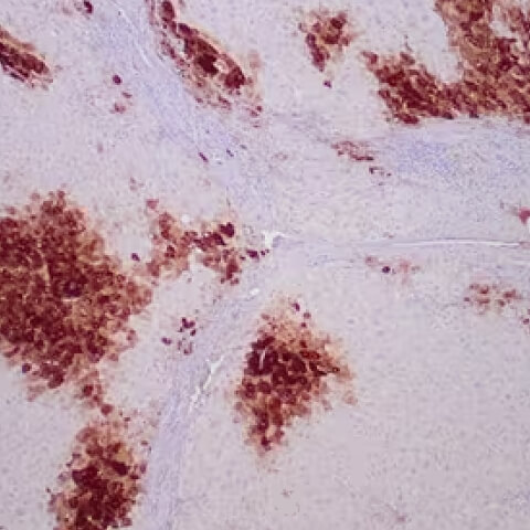
Anti-Vimentin Antibody [VI-10] Cross-Adsorbed
Recombinant mouse monoclonal antibody to Cytokeratin 18
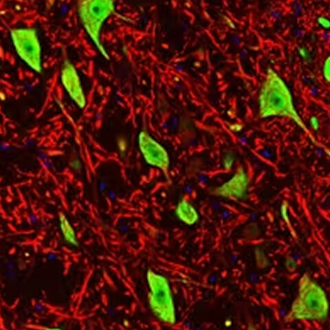
Anti-Human Nuclear Antigen Antibody
Recombinant mouse monoclonal antibody to Cytokeratin 18

Alternative products to anti-GFAP antibody (A85419)
Goat Anti-Rabbit IgG H&L Antibody (FITC), Cross
Recombinant mouse monoclonal antibody to Cytokeratin 18
Anti-Vimentin Antibody [VI-10] Cross-Adsorbed
Recombinant mouse monoclonal antibody to Cytokeratin 18
Anti-Human Nuclear Antigen Antibody
Recombinant mouse monoclonal antibody to Cytokeratin 18
Proteins predicted to interact with GFAP
Predicted protein interactions based upon String database. Revelancy score correlates with probability of interaction.
eEF1A1 antibodies
95.6% relevancy score
NeuN antibodies
93.1% relevancy score
S100 beta antibodies
92.5% relevancy score
Olig2 antibodies
91.9% relevancy score
Iba1 antibodies
91.3% relevancy score
Nestin antibodies
88.6% relevancy score
EAAT2 antibodies
88.5% relevancy score
CNPase antibodies
88.3% relevancy score
Myelin basic protein antibodies
87.6% relevancy score
S100 beta antibodies
87.3% relevancy score